| Human vertebral body; analysis of facet
joint pressure |
|
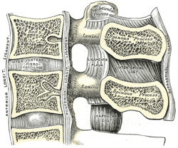 |
| Fig. 1: Sagittal
section throught the human lumbar vertebral spine. Facet
joints are shown right. The ligaments shown are intertransverse
ligament (ITL), supraspinous ligament (SSL), interspinous
ligament (ISL), ligamentum flavum (LF), anterior longitudinal
ligament (ALL), posterior longitudinal ligament (PLL)
and capsular ligament (CL). |
The curvature of vertebral facet joints may
play an important role in the study of load bearing characteristics
and clinical interventions such as graded facetectomy. In
previously published finite element simulations of this procedure,
the curvature was either neglected or approximated with a
varying degree of accuracy. Here we study the effect of the
curvature in three different load situations by using a numerical
model which is able to represent the actual curvature without
any loss of accuracy. The results show that previously used
approximations of the curvature lead to good results in the
analysis of sagittal moment/rotation. However, for sagittal
shear-force/displacement and for the contact stress distribution,
previous results deviate significantly from our results. These
findings are supported through related convergence studies.
Hence we can conclude that in order to obtain reliable results
for the analysis of sagittal shear-force/displacement and
the contact stress distribution in the facet joint, the curvature
must not be neglected. This is of particular importance for
the numerical simulation of the spine, which may lead to improved
diagnostics, effective surgical planning and intervention.
The proposed method may represent a more reliable basis to
optimize the biomedical engineering design for tissue engineering
or, for example, for spinal implants.
|
|
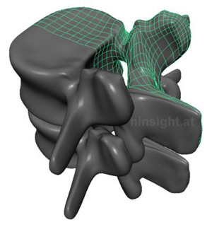 |
| Fig. 2: Contact interaction between
lumbar vertebral bodies L4 and L5. Representation of both
bodies by means of subdivision surfaces. The ligaments
and the intervertebral disc are not show, |
The geometry of the human vertebrae, and,
in particular, the curvature of the facet joints, is highly
complex. For our study we have not achieved the desired accuracy
with CT-image-based analyses. Hence, we used slices from the
Visible-Human-Data project [1]. The slices were available
in distances of 1 mm. The accurate consideration of the facet
geometry in the finite element simulation requires a special
technique for mesh generation and surface description. Therefore,
the geometry of L2 to L5 was traced with so called subdivision
surfaces [2]. They help to model smooth biological surfaces
up to any level of accuracy, similarly to NURBS, Bézier
or Hermite splines. However, their advantage is that they
can deal with arbitrary mesh topologies, i.e. more or less
than four quadrilaterals can meet in one node. Such a geometrical
situation is encountered quite frequently, and can not be
treated easily with NURBS, Bézier or Hermite splines.
In contrast, previous studies represented the facet joints
as planar surfaces, which are described by their angular alignment
in space ([3], [4], [5]). However, this approach has the major
drawback of not being able to accurately describe the facet
surface (which is not planar). This is now possible with the
presented novel approach by the use of subdivision surfaces.
We do not present the detailed data set associated with the
subdivision surface description of the facet joints here;
instead we refer to the Visible Human Project [1], from which
our model geometry is originated. The gap size was 0.4 mm
for all motion segments.
|
| Contact stress distribution |
|
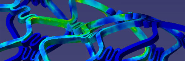
| The motion segment L4-L5, represented
by subdivision surfaces, is shown in Figure 3(a). Here we
used, in particular, the Catmull-Clark subdivision surface
[2], which offers C2-continuity in the regular mesh domain
and C1-continuity at irregular nodes. The contact stress distribution
along the white dashed path starting from the inferior position
`A' (see Fig. 3(a)) is plotted in Fig. 3(b). Therein, we compare
the results for piecewise planar contact elements and for
subdivision surfaces, both for different mesh densities.
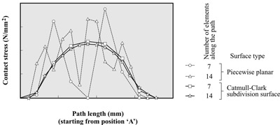 |
| Fig. 3: Contact
stress distribution along a vertical path between the
facet joints for different mesh densitites and contact
surface continuities. |
|
|
1. The NPAC visible human visualization project (1995)
http://rockefeller.univ-lyon1.fr/VisibleHumanProjectEnglish/VisibleHuman.html,
Norhteast Parallel Architecture Center, Syracuse University
2 . Catmull E, Clark J (1987) Recursively generated B-spline
surfaces on arbitrary topological meshes. Comput Aided Design
10:350-355
3. Natarajan RN, Williams JR, Andersson GB (2003) Finite element
model of a lumbar spinal motion segment to predict circadian
variation in stature. Comput & Structures 81:835-842.
4. Sharma M, Langrana NA, Rodriguez J (1995) Role of ligaments
and facets in lumbar spine stability. Spine 20:887-900
5. Sharma M, Langrana NA, Rodriguez J (1998) Modeling of facet
articulation as a nonlinear moving contact problem: senstivity
study on lumbar facet response. J Biomech Engr 120:118-125
|